CARDIOVASCULAR ASSESSMENT
ANATOMY AND PHYSIOLOGY UPDATE
The Thorax
The first part of this course is an update of the anatomical structures and the physiology of the cardiovascular system in relationship to the nursing care of the patient with a related disorder. If you need to review the related structures and/or function which we will be discussing, please refer to any basic anatomy textbook; one of which you probably have hidden away somewhere in your closet or garage since nursing school. However, it is not required that you use any other reference. All information needed to pass the test at the end of this course, will be included in the text.
We will list the most important structures of the thorax and entire CV system only so that you can relate these to the clinical approach that we will use. Following is an illustration of the thorax and the heart in relationship to other structures which will be noticed upon visual examination. The thorax has a characteristic shape, size and movement.
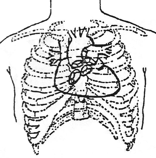
Thorax – Be sure you can identify the following:
- Sternum – mid chest, flat, non-protruding
- Ribs – slope of ribs, intercostal spaces, costal margins
- Heart – heartbeat in some cases, can be visible as a pulsation in the thorax at lower costal margin
- Shoulder – patient’s shoulder should be relaxed, and at a 90 degree angle to the patient – look for abnormal angles and musculature which might indicate overuse of accessory muscles
- Neck veins – should not normally be visible
- Clavicle – clavicular line horizontal with no protrusions during breathing
- Respirations – normal respiration should be unlabored and comfortable
The Heart
Gross structures
- Musculature-pericardium, fibrous & serous epicardium, visceral serous pericardium, myocardium, heart muscle
- Muscle cell (microscopic structures), central nucleus, sarcoplasm, sarcolemma, sarcomere-the contractile unit, intercalated discs
- Pattern of blood flow through the structures of the heart; atria, ventricles, valves
Chambers
Right side of Heart
- Right atrium – the thin-walled atrium, low relative pressure receives blood from superior and inferior vena cavae, the coronary sinus and thebesian veins. The outflow of blood through tricuspid valve.
- Right ventricle – relatively thin muscle wall, crescent-shaped, papillary muscles, chordae tendineae, low pressure. Outflow through the pulmonic valve to the pulmonary artery.
Left side of the heart
- Left atrium, - thicker muscle, medium pressure of blood, inflow of blood through the four pulmonary veins. Outflow is through the mitral valve.
- Left ventricle – largest muscle mass, high pressure blood flow, papillary muscles, spring-like pump action. Outflow of blood through the aortic valve and the aorta.
Heart Valves
Atrioventricular Valves:
- Tricuspid – has three leaflets, controlled by papillary muscles; chordate tendineae
- Mitral valve – two cusps, controlled by papillary muscles and the chordae tendineae
Semilunar Valves:
- Pulmonic valve – three-leaflet valve, formed by fibrous ring, tendinous tubercle midpoint free edges
- Aortic valve – three leaflets, also formed by fibrous ring, tendinous tubercle midpoint free edges.
Vasculature of the Heart
- Right coronary artery – most branches of this artery anastomose distally with left anterior descending.
- Left coronary artery – divides into two main branches, left ant. descending and left circumflex artery.
- Great cardiac vein – largest system, forms coronary sinus, drains left ventricle primarily.
- Anterior cardiac veins – empty directly into right atrium.
- Thebesian veins – smallest system, empty into right atrium
Conduction System of the Heart
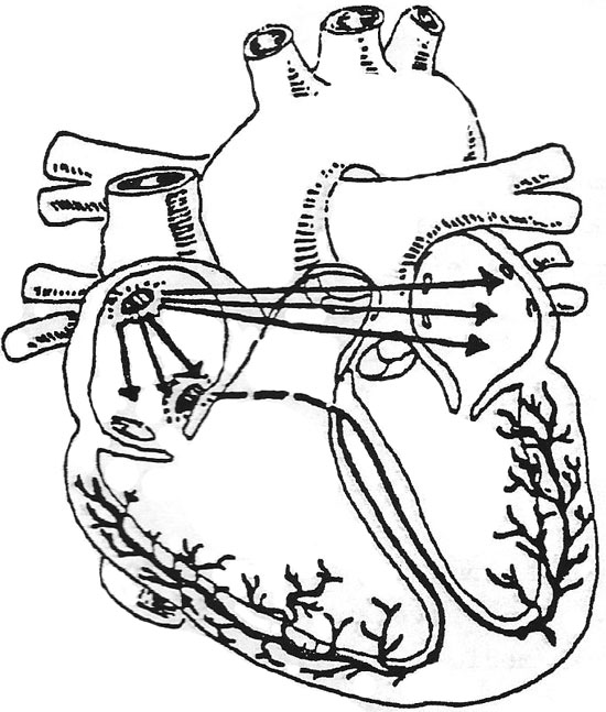
- SA (Sino-atrial node)
- Atrial preferential pathways – anter, internodal, middle, posterior internodal
- AV (Atrio-ventricular node)
- Bundle of HIS
- Left Bundle Branch
- Right Bundle Branch
- Purkinje fibres
Contractility of Heart Muscle
At this time it is appropriate to mention the physiology of muscle contractility. Electrical conduction in the heart is unique and remarkable.
Heart muscle possesses the following properties:
- Automaticity- pacemaker ability
- Conductivity-each cell has the ability to conduct impulses
- Contractility-ability to contract (make each cell shorter or longer)
- Irritability-each cell has ability to contract on its own, to send impulses to cells without it first being stimulated from another source
These properties make the myocardium different from other muscle cells in the body. The normal activity of the heart conducts impulses from one point (SA node) to another point (individual cells), thus stimulating a uniform and effective contraction.
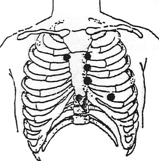
Various factors affect the activity of cardiac muscle. The availability of oxygen, afterload, nervous control, muscle condition and other factors can affect the force of the contraction and affects blood flow through the heart. Drugs can also affect the contraction of the heart. The nurse should be aware of all the factors which influence heart activity and heart sounds. The below figure shows points on the thorax where heart sounds are best heard. Later in this text, these points on the thorax and heart sounds will be discussed in greater detail.
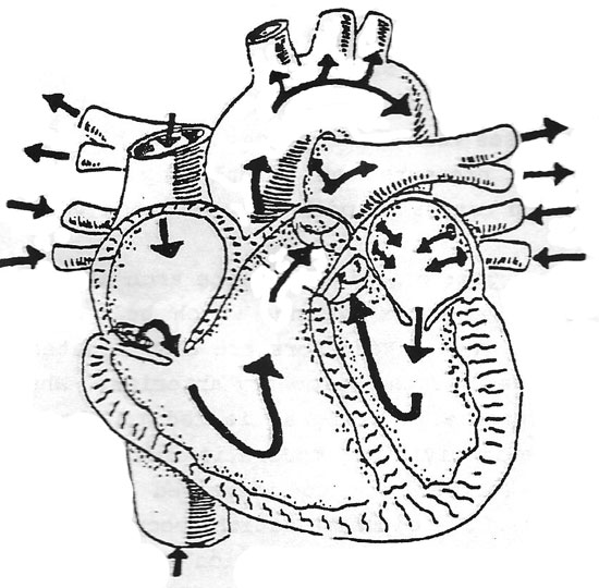
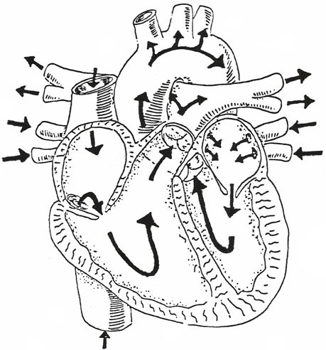
Refer to the figure below to view blood flow through the heart. Blood is shown as it enters the heart, circulates and then leaves the heart. In relation to the physical assessment performed by most nurses, keep in mind the changes in circulation which will be assessed. Impeded flow may cause extra heart sounds and/or physical changes. Also, reduced flow will usually cause changes that can be assessed by the nurse.
Physical characteristics important to blood flow:
- Diameter of the blood vessels
- Cross-section areas of the chambers and vessels
- Length of the vessels
Quantities of blood:
- Heart 18%
- Pulmonary vessels 12%
- Large arteries 8%
- Small arteries 5%
- Arterioles 2%
- Capillaries 5%
- Small veins 25%
- Large veins 25%
Velocities of blood flow:
The velocity of blood flow is directly related to the amount of circulating blood volume and the area of the vessels. Blood returns to the heart from the general circulation. Almost 50% of all blood in the body is in the systemic veins of the body. This includes small veins and venules and blood in the pulmonary circulation. The small veins usually offer little resistance to blood flow. The large veins do offer much resistance to the flow of blood to the heart. This is an important nursing implication, as the patient who is more active will have better flow of blood back to the heart.
With reduced activity, the blood tends to pool in the large vessels and can lead to severe venous stasis. Blood returns to the heart via the superior and inferior vena cavae and into the right atrium.
From the right atrium blood flows to the right ventricle and is then propelled into pulmonary circulation. After blood is aerated with fresh oxygen, it is returned to the left side of the heart into the left atrium.
From the left atrium the blood is ejected into the left ventricle. The left ventricle then pumps the blood out of the heart into the general circulation. The aorta is the first vessel to carry blood, and, at the same time, the coronary arteries are being fed oxygenated blood to circulate through the heart.
The above is only a brief outline of the circulation of blood. Be sure you can trace the blood through the heart. Be sure that you can name all the valves and chambers of the heart as blood flows through. You should also be able to list the major arteries of the body. Later, when performing the assessment, it will be necessary for you to know these vessels and their location.
Next: Cardiovascular Assessment Continued